

시설 및 장비 소개
Shared Research Facility
Department of Brain and Cognitive Sciences
보유 장비
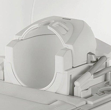
MAGNETOM Trio, A Tim System 3T
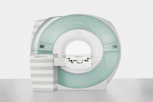
- 1. Actively Shielded Magnet System
-
- 1) Field Strength: 3 Tesla
- 2) Magnet Homogeneity: Typical
- 10cm DSV: 0.001 ppm or less
- 20cm DSV: 0.01 ppm or less
- 30cm DSV: 0.05 ppm or less
- 40cm DSV: 0.5 ppm or less
- 50cm DSV: 1.2 ppm or less
- 2. Advanced High Order Shim System
- 3. Gradient System
-
- 1) Gradient Amplitude: max. 45mT/m or more
- 2) Slew Rate: 200 mT/m/sec or more
- 3) Max. b-value: 10,000 or more
- 4. Digital RF System
- 5. Coil Technology
-
-
Drag from side to side.
1) Head Matrix 12 Ch. Coil or 8 Ch. SENSE Head Coil 2) Neck Matrix 4 Ch. Coil or 16 Ch. SENSE NV Coil 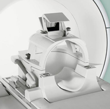
3) Spine Matrix 24 Ch. Coil or 33 element SENSE Spine Coil 4) 32 Ch. Head Coil approved from FDA as of 2009 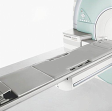
5) animal coil 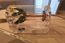
-
- 6. Patient Handling Unit
-
- 1) Provided patient table
- 2) Dual-way intercom system
- 3) Automated voice commands (e.g. for breath-hold commands)
- 4) Physiological Measurement Unit (Pulse oximeter, ECG & Respiratory Triggering)
- 5) Patient monitoring camera
- 7. Standard and Advanced Software Package
-
- Advanced software package should include the methods for parallel imaging, functional imaging, diffusion
- tensor imaging (256-direction or more), spectroscopic imaging, chemical shift imaging & Motion correction s/w
- 8. fMRI Trigger Converter
-
- With the "fMRI Trigger Converter this signal can be converted to an electrical signal
(TTL/BNC and RS 232 interface for PC: modes: toggle or impulse)
- With the "fMRI Trigger Converter this signal can be converted to an electrical signal
- 9. Remote Planning and Processing Software
-
- syngo Expert-I or Portal
SIEMENS Neuro 3-D
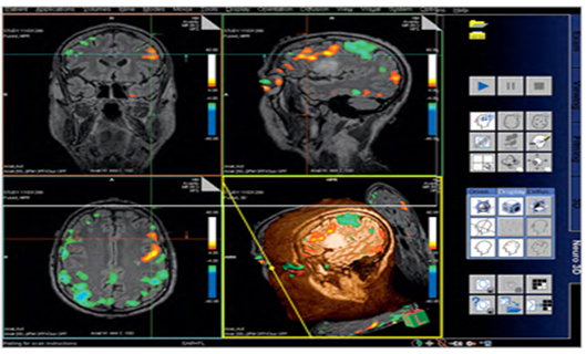
Many new tools are available for imaging the brain for surgical planning. Neuro 3D in the lastest syngo MR B15 software provides a comprehensive diagnostic tool that utilizes cutting-edge imaging techniques such as BOLD (Blood Oxygen Level Dependent) and Diffusion Tensor Imaging (DTI). These imaging techniques have been mostly research in the past, but now they are clinical reality with new imaging te chniques and post processing BOLD imaging with DTI was performed on a patient with a known lesion inthe left hemisphere to assess a surgicalapproach. The neurosurgeon was concerned that damage would be done tothe patients motor skills.
The first important imaging technique is a 3D sequence loaded into Neuro 3D. In this case, a SPACE DarkFluid sequence was used to define tumor borders. syngo SPACE provides high isotropic resolution in a very short period of time using iPAT (integrated Parallel Acquisition Techniques). Next the GLM (General Linear Model) was loaded into Neuro 3D that was acquired from our 3D PACE epi bold sequence. The GLM was calculated inline with Inline BOLD imaging, so no post processing was needed to get the GLM. Next, the tensor data was loaded into Neuro 3D. This too was calculated inline, with Inline diffusion
E-PRIME3.0

Extensions for fMRI
E-Prime® Extensions for fMRI (EEfMRI) software is designed to optimize E-Prime® experiments for fMRI research. EEfMRI allows you to synchronize the start of your experiment with the first scanner trigger pulse along with several valuable features to enhance the control you have over your experiment.
Implementing EEfMRI into your current experiments is achieved by simply dragging and dropping the correct EEfMRI package calls into the E-Prime® experiment in the appropriate places. EEfMRI is designed to integrate with other PST hardware and software to increase usability for researchers while maintaining the millisecond accuracy of E-Prime®.
Features
Synchronization of the start of the experiment with the scanner trigger pulse
Simplified logging of user-specified experimental events to tailor behavioral output for later analysis.
Easy to implement menu system that allows the experimenter to group multiple tasks into a single experiment and interactively choose which tasks to present at run time
Ability to interrupt a running task in a controlled manner to restart tasks without terminating the entire experiment.
New documentation including samples and tutorials
Turnkey fMRI block sample (MapperOne.es2) based on a published experiment* that reliably activates important functional areas
BrainVoyager
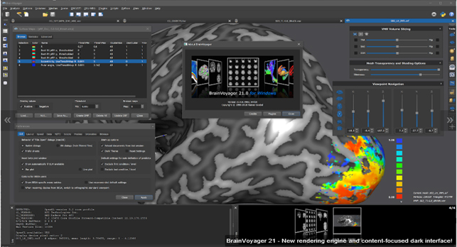
Our flagship product BrainVoyager is a powerful neuroimaging software package for data management and data analysis. It started as a tool for the analysis of anatomical and functional MRI data sets but has evolved over the years into a multi-modal analysis tool for fMRI, DTI, EEG and MEG data. The software is highly optimized and user friendly running on all major computer platforms; the current version runs on Windows (7/8/10), Linux (e.g. Ubuntu, SUSE, Fedora) and macOS (10.10 or higher).
BrainVoyager is a 64 bit program supporting analyses of large data sets that need more than 3 GB of RAM. In order to obtain maximum speed on each platform, BrainVoyager has been programmed in C++ with optimized and highly efficient statistical, numerical, and image processing routines. It supports on all platforms fast parallelized basic math routines using the Intel Math Kernel Library (MKL). The software also exploits modern multi-core, multi-processor hardware for the most demanding computational routines. Multiple parallel processing pipelines of modern graphic cards (GPU's) are used for real-time volume rendering, data filtering and sinc interpolation. The surface rendering environment ("surface module") has been implemented using OpenGL.
The interactive graphical user interface (GUI) has been built using the award-winning cross-platform Qt C++/QML toolkit from Digia (formerly Nokia and Trolltech). Using cross-platform C++/QML code for all aspects of the program, BrainVoyager provides a native and responsive user interface and powerful computational routines on all supported platforms.
Eyetracker(EyeLink 1000)
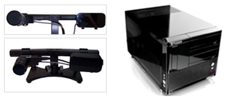
The EyeLink 1000 Plus is the world’s most precise and accurate video-based eye tracker, sampling binocularly at up to 2000 Hz. It is also the most flexible and customizable
eye tracker, with multiple mounting options, interchangeable lenses, and multiple head free-to-move remote configurations.
The EyeLink 1000 Plus eye tracker has outstanding precision and accuracy with low spatial noise and high sampling rates, in both head supported and head free-to-move Remote Mode.
Choose from a range of camera mounting options – Desktop Mount, Tower Mount, Arm Mount, and Primate Mount.
Fast, accurate and reliable eye tracking across a wide range of populations – from infants through to elderly participants and even non-human species.
The EyeLink 1000 Plus smoothly integrates with SR Research Experiment Builder, Data Viewer, WebLink, and third-party software and tools including E-Prime, Presentation, MATLAB and Psychtoolbox, etc.
Audio system(BOLDfonic)
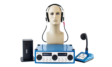
BOLDfonic delivers high-fidelity acoustic stimuli for fMRI while also attenuating scanner noise. Our electro-dynamic driver technology uses the magnetic field of the MRI scanner to drive the headphone membranes. This results in powerful speakers with an excellent frequency response across a wide dynamic range. A novel USB sound processor with automated synchronous triggering capabilities and a fully–loaded amplifier system provide all the controls you need for rigorous multimodal EEG/fMRI studies.
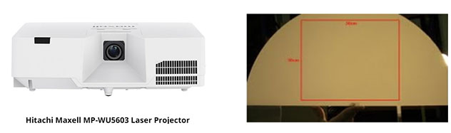
Visual display system (Projector / Screen)
-
Drag from side to side.
| 모델명 | MP-WU5603 | 디스플레이 | 0.64"3LCD |
|---|---|---|---|
| 밝기 | 6,000 lm(루멘) | 명암비 | 1,500,000:1 |
| 해상도 | WUXGA 1920x1200 | 화면비 | 16:10 |
| 화면사이즈 | 30 ~ 300" | 광원타입 | Laser Phosphor |
| 광원 수명 | 20,000 시간 | 렌즈쉬프트 | 상하 +56.5% / 좌우 ±4.6% |
| 키스톤 | 상하 ±30° / 좌우 ±30° | 종횡왜곡보정 | 퍼펙트 핏 |
| 줌/포커스 | 수동/자동 | 투사거리비율 | 1.4-2.4:1 |
| 입력단자 | 2 X HDML | 출력단자 | 1 X 15pin D-sub(IN2 RGB) |
| 2 X 15pin D-sub(RGB) | 1 X Φ3.5 jack Audio | ||
| 1 X RCA(Composite) | 컨트롤/서비스단자 | 1 X HDBaseT | |
| 1 X 2RCA(L/R) Audio | 1 X RS-232C | ||
| 2 X Φ3.5 jack Audio | 1 X RJ-45 | ||
| 스피커 | 16W | 1 X USB(A) | |
| 비디오 포맷 | NTSC, NTSC4.43, PAL, PAL-M, -N, SECAM, 480i/p, 576i/p, 720p, 1080i/p, 4K(3840x2160p@24/25/30,4096x2160p@24) | 작동온도 | 0 ~ 40℃ |
| 정격전압 | AC 100~240V, 50/60 Hz | 소비전력 | 400W |
| 외형크기(WxHxD) | 506 x 136 x 424 mm | 무게 | 8.5kg |
Response key & Interface
4 Button Bimanual
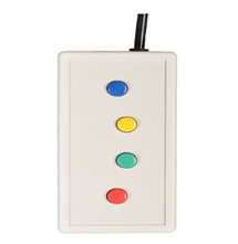
Current Designs' fMRI response pad is the perfect choice for computer response tasks used in the human brain mapping project. It is non-magnetic, non-electronic, and entirely made out of plastic, so will not add noise to the images or raise safety concerns in the MR/MEG room or similarly demanding environments. There is a less expensive, non-MR trainer available for this response pad for use outside the magnet room. Note: Only one response device can be connected to an interface at a time. There is a two-handed version of this response device. If a bimanual devices is needed order HHSC-2x4-C instead of HHSC-1x4-CR and HHSC-1x4-CL.
8 Button Bimanual Straight Lines
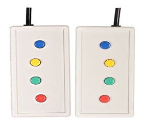
Current Designs' fMRI response pad is the perfect choice for computer response tasks used in the human brain mapping project. It is non-magnetic, non-electronic, and entirely made out of plastic, so will not add noise to the images or raise safety concerns in the MR/MEG room or similarly demanding environments. There are less expensive non-MR trainers (TR-1x4-L) available for this response pad for use outside the magnet room.
Pyka 8 Button Bimanual - Thumbs Disabled
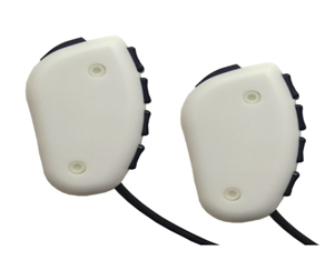
Current Designs' new fMRI eight button response pad is the perfect choice for a wide range of computer response tasks used in the human brain mapping project. It is non-magnetic, non-electronic, and entirely made out of plastic, so will not add noise to the images or raise safety concerns in the MR/MEG room or similarly demanding environments.
Tethyx
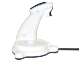
Current Designs' fMRI joystick is the perfect choice for a wide range of computer response tasks used in the human brain mapping project. It is non-magnetic, non-electronic, and entirely made out of plastic, so will not add noise to the images or raise safety concerns in the MR/MEG room or similarly demanding environments. There is a less expensive, non-MR trainer available for this response pad for use outside the magnet room.
Dial
Description Current Designs' new fMRI dial response pad is the perfect choice for a wide range of computer response tasks used in the human brain mapping project. It is non-magnetic, non-electronic, and entirely made out of plastic, so will not add noise to the images or raise safety concerns in the MR/MEG room or similarly demanding environments.
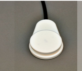
932Interface
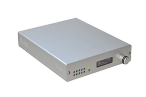
The opto-electronic interface receives optical signals from the handheld devices in the MR suite, and converts them into electronic signals for the computer. This is our top of the line model.
In keeping with our earlier interface units, the 932 is standards-based: the outputs are not designed to be used with a specific program or hardware platform but are instead designed to work with everything. This interface provides complete USB, Serial and Parallel outputs.
904 Interface
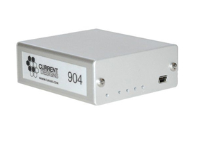
The opto-electronic interface receives optical signals from the handheld devices in the MR suite, and converts them into electronic signals for the computer. This is our top of the line model.
In keeping with our earlier interface units, the 932 is standards-based: the outputs are not designed to be used with a specific program or hardware platform but are instead designed to work with everything. This interface provides complete USB, Serial and Parallel outputs.
8 Fiber Bundle 80 ft

8 fiber removable fiber optic bundle, 80 ft (25 m) long
This bundle has been replaced by the Bundle Extension
Physiological system
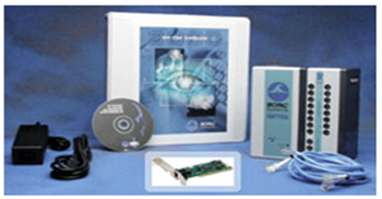
- 1. EDA100C-MRI(전극활성증폭기) - Electrodermal response amplifier for MRI environment
-
- 1) MECMRI-TRANS - MRI Components for Transducer Amplifiers
- 2) TSD203 - Electrodermal response transducer
- 3) GEL101 - Isotonic Electrode paste
- 2. PPG100C-MRI - Pulse plethysomogram amplifier for MRI environment
-
- 1) MECMRI-TRANS - MRI Components for Transducer Amplifiers
- 2) TSD200-MRI - Photo Plethysmograph for PPG100C-MRI
- 3. ECG100C-MRI(심전도) - Electrocardiogram amplifier for MRI environment
-
- 1) MECMRI-BIOP - MRI Components for Biopotential Amplifier
- 2) LEAD108A - Lead, 3.6m MRI-compatible, for EL508
- 3) Electrode, Disp,MRI-compatible(pkg.30)
TMS
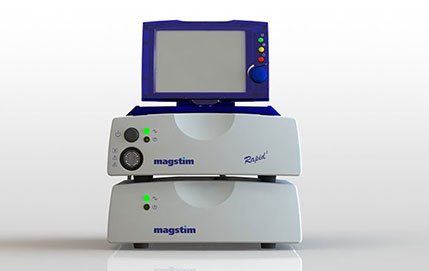
The Magstim Rapid2 is capable of high-frequency repetitive protocols for both cortical and peripheral stimulation. With a package to fit a wide variety of research purposes, the Magstim Rapid² has been used in research studies worldwide.
NAS
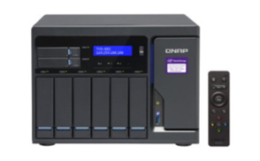
QNAP® Systems, Inc. today launched the new TVS-x72N 5GbE NAS series, powered by 8th Generation Intel® Core™ i3 processors and features a 5GBASE-T port to deliver transfer speeds up to five times faster than Gigabit Ethernet, making 4K video transfer, display, and editing smooth and streamlined. The TVS-x72N comes with two built-in M.2 SSD slots for enabling SSD caching, features HBS 3 with data deduplication for faster data backup and recovery, and provides cloud storage gateway functions to create a cost-effective hybrid cloud storage environment.
“With computers and networking devices now widely supporting Wi-Fi 6 (802.11ax), the common Ethernet networking speed has increased from 1 Gigabit (GbE) to 2.5 GbE and 5 GbE,” said David Tsao, Product Manager of QNAP, adding, “Users can add the TVS-x72N 5GbE NAS that offers four transfer speeds to their network environment to optimize their network storage usage experience.”
주소 : (우)0882 서울특별시 관악구 관악로 1(신림동)
상호 : 서울대학교 자연과학대학 뇌인지과학과 공동기기실 대표자명 : 유재준 사업자등록번호 : 119-82-11049 연락처 : +82-2-880-4999메일 : snu.bcs.mri@gmail.com
Copyrightⓒ 2020 ICMRC All Rights Reserved.